

Tip localization of an atomic force microscope in transmission microscopy with nanoscale precision
20-Mar-2015
Rev. Sci. Instrum., 86, 035109, http://dx.doi.org/10.1063/1.4915145
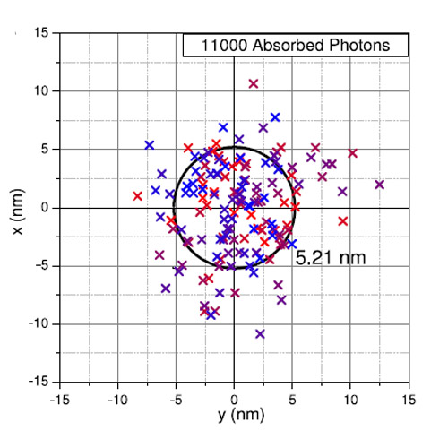
Since the atomic force microscope (AFM) has evolved into a general purpose platform for mechanical experiments at the nanoscale, the need for a simple and generally applicable localization of the AFM cantilever in the reference frame of an optical microscope has grown. Molecular manipulations like in single molecule cut and paste or force spectroscopy as well as tip mediated nanolithography are prominent examples for the broad variety of applications implemented to date. In contrast to the different kinds of superresolution microscopy where fluorescence is used to localize the emitter, we, here, employ the absorbance of the tip to localize its position in transmission microscopy. We show that in a low aperture illumination, the tip causes a significant reduction of the intensity in the image plane of the microscope objective when it is closer than a few hundred nm. By independently varying the z-position of the sample slide, we could verify that this diffraction limited image of the tip is not caused by a near field effect but is rather caused by the absorbance of the transmitted light in the low apex needle-like tip. We localized the centroid position of this tip image with a precision of better than 6 nm and used it in a feedback loop to position the tip into nano-apertures of 110 nm radius. Single-molecule force spectroscopy traces on the unfolding of individual green fluorescent proteins within the nano-apertures showed that their center positions were repeatedly approached with very high fidelity leaving the specific handle chemistry on the tip’s surface unimpaired