

Visualization and targeted disruption of protein interactions in living cells
24-Oct-2013
Nature Communications, 2013, doi:10.1038/ncomms3660, 4, Article number: 2660 published on 24.10.2013
Nature Communications, online article
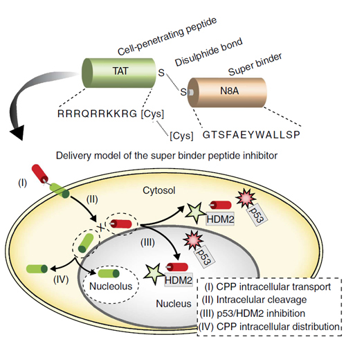
Protein–protein interactions are the basis of all processes in living cells, but most studies of these interactions rely on biochemical in vitro assays. Here we present a simple and versatile fluorescent-three-hybrid (F3H) strategy to visualize and target protein–protein interactions. A high-affinity nanobody anchors a GFP-fusion protein of interest at a defined cellular structure and the enrichment of red-labelled interacting proteins is measured at these sites. With this approach, we visualize the p53–HDM2 interaction in living cells and directly monitor the disruption of this interaction by Nutlin 3, a drug developed to boost p53 activity in cancer therapy. We further use this approach to develop a cell-permeable vector that releases a highly specific peptide disrupting the p53 and HDM2 interaction. The availability of multiple anchor sites and the simple optical readout of this nanobody-based capture assay enable systematic and versatile analyses of protein–protein interactions in practically any cell type and species.