

Rapid time course of action potentials in spines and remote dendrites of mouse visual cortex neurons
15-Feb-2010
The Journal of Physiology, 2010, doi: 10.1113/jphysiol.2009.184960 published on 15.02.2010
The Journal of Physiology, online article
The Journal of Physiology, online article
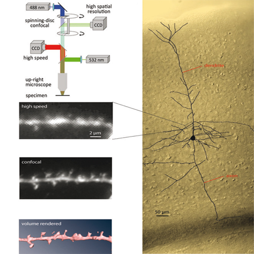
Axonally initiated action potentials back-propagate into spiny dendrites of central mammalian neurons and thereby regulate plasticity at excitatory synapses on individual spines as well as linear and supralinear integration of synaptic inputs along dendritic branches. Thus, the electrical behavior of individual dendritic spines and terminal dendritic branches is critical for the integrative function of nerve cells. The actual dynamics of action potentials in spines and terminal branches, however, are not entirely clear, mostly because electrode recording from such small structures is not feasible. Additionally, the available membrane potential imaging techniques are limited in their sensitivity and require substantial signal averaging for the detection of electrical events at the spatial scale of individual spines. We made a critical improvement in voltage-sensitive dye imaging technique to achieve multisite recordings of backpropagating action potentials from individual dendritic spines at a high frame rate. With this approach, we obtained direct evidence that in layer 5 pyramidal neurons from the visual cortex of juvenile mice the rapid time-course of somatic action potentials is preserved throughout all cellular compartments, including dendritic spines and terminal branches of basal and apical dendrites. The rapid time-course of the action potential in spines may be a critical determinant for the precise regulation of spike-timing dependent synaptic plasticity within a narrow time window.