

αβ T-cell receptors from multiple sclerosis brain lesions show MAIT cell–related features
07-May-2015
Neurology, 2015, doi: 10.1212/NXI.0000000000000107, published on 07.05.2015
Neurology, online article
Neurology, online article
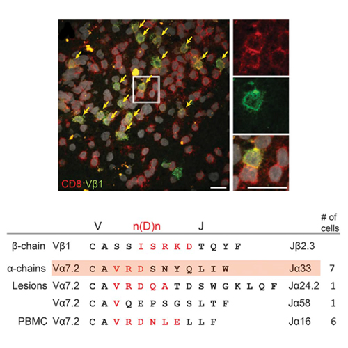
Objectives: To characterize phenotypes of T cells that accumulated in multiple sclerosis (MS) lesions, to compare the lesional T-cell receptor (TCR) repertoire of T-cell subsets to peripheral blood, and to identify paired α and β chains from single CD8+ T cells from an index patient who we followed for 18 years. Methods: We combined immunohistochemistry, laser microdissection, and single-cell multiplex PCR to characterize T-cell subtypes and identify paired TCRα and TCRβ chains from individual brain-infiltrating T cells in frozen brain sections. The lesional and peripheral TCR repertoires were analyzed by pyrosequencing. Results: We found that a TCR Vβ1+ T-cell population that was strikingly expanded in active brain lesions at clinical onset comprises several subclones expressing distinct yet closely related Vα7.2+ α chains, including a canonical Vα7.2-Jα33 chain of mucosal-associated invariant T (MAIT) cells. Three other α chains bear striking similarities in their antigen-recognizing, hypervariable complementarity determining region 3. Longitudinal repertoire studies revealed that the TCR chains that were massively expanded in brain at onset persisted for several years in blood or CSF but subsequently disappeared except for the canonical Vα7.2+ MAIT cell and a few other TCR sequences that were still detectable in blood after 18 years. Conclusions: Our observation that a massively expanded TCR Vβ1-Jβ2.3 chain paired with distinct yet closely related canonical or atypical MAIT cell–related α chains strongly points to an antigen-driven process in early active MS brain lesions.